In XRAY, the earliest changes are seen in adjacent soft tissues +/- muscle outlines with swelling and loss or blurring of normal fat planes. An effusion may be seen in an adjacent joint.
In general, osteomyelitis must extend at least 1 cm and compromise 30 to 50% of bone mineral content to produce noticeable changes on plain radiographs.
Look for:
regional osteopenia
periosteal reaction/thickening (periostitis): variable; may appear aggressive, including the formation of a Codman’s triangle
focal bony lysis or cortical loss
endosteal scalloping
loss of trabecular bone architecture
new bone apposition
eventual peripheral sclerosis
In chronic or untreated cases, the eventual formation of a sequestrum, involucrum, and/or cloaca
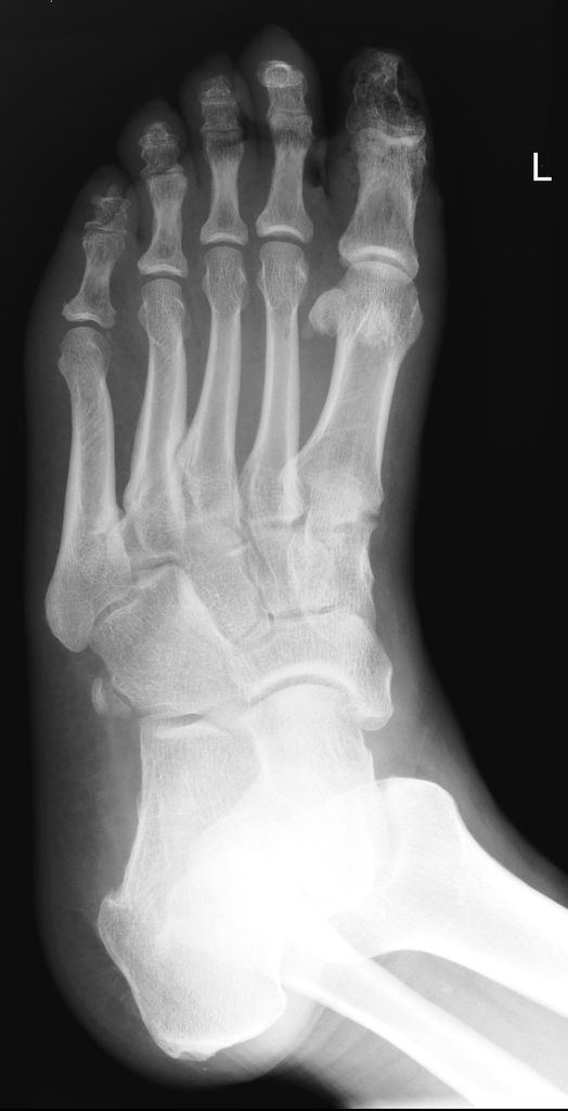
This radiographs shows typical findings seen very commonly in patients with diabetic foot ulcer.
There is loss of soft tissue over the great toe, with further lucencies in the surrounding soft tissue. This is very suggestive of superimposed infection. The underlying phalanx shows patchy osteoporosis and is suggestive of osteomyelitis.
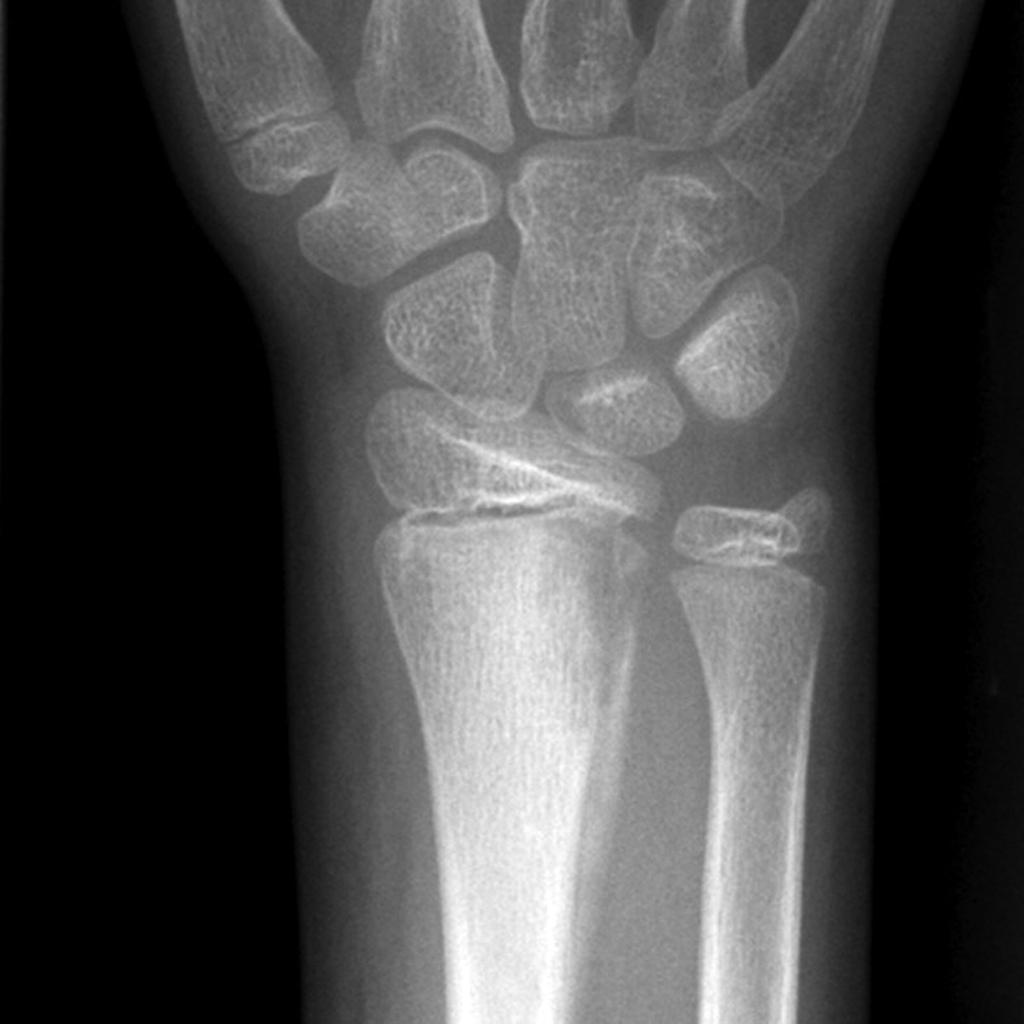
12 year old with osteomyelitis of the distal radius